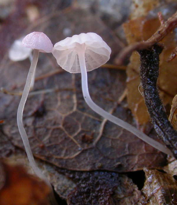
Typically on fallen, decaying leaves of
Quercus. Widely distributed. Autumn. In Norway fairly common in Vestfold and Agder. Records on Betula and Salix leaves in other parts of the country have proved to be Mycena exilis.
Pileus 1-5 mm
across, hemispherical to parabolical, becoming plano-convex, often with somewhat
flattened centre, or sometimes with a small umbo, sulcate,
translucent-striate, pruinose, glabrescent, pale pink, pinkish-apricot
to brownish-pink, often darker at the centre, with age often
paler to whitish pink, sometimes almost entirely white. Lamellae
5-13 reaching the stipe, ascending to subhorizontal, fairly
broad, narrowly to broadly adnate, sometimes decurrent with
a short tooth, pink to pinkish-white, the edge convex, white.
Stipe 5-15(-40) x 0.1-0.4 mm,
straight to curved, equal, pruinose, glabrescent except
for the base, the base somewhat bulbous, fairly dark grey
at the apex in younger stages and grey below, becoming watery
grey to watery white, insitituous or sometimes attached with a whorl of very thin, radiating fibrils. Odour
none. Taste not recorded.
Basidia
19-25 x 6-9 µm, slender-clavate, 2-spored, with
sterigmata up to 8 µm long. Spores
10-12.5 x 3.8-5 µm, Q 1.9-2.5, Qav ≈ 2.1, elongated pip-shaped to somewhat
cylindrical, amyloid. Cheilocystidia
14-29 x 6-16 µm, forming a sterile band, clavate to
obpyriform, covered with fairly numerous, evenly
spaced, cylindrical excrescences 1-5 µm long.
Pleurocystidia absent. Lamellar trama dextrinoid, vinescent in Melzer’s reagent. Hyphae
of the pileipellis 3.5-12 µm wide,
densely covered with warts, terminal cells up to 52 x 14 µm, inflated.
Hyphae of the cortical layer of the stipe
2-3 µm wide, diverticulate, with excrescences 1.5-5 x 0.5-1.5 µm. Caulocystidia
clavate to subglobose, diverticulate. Clamp connections absent.
Similar-looking specimens found on fallen leaves of Betula in mountain birch forest and on fallen leaves of Salix in alpine areas have been associated with this species, but they should be referred to the recently described M. exilis (Aronsen & Gulden 2007). It differs in having 4-spored basidia, clamps, somewhat smaller but slightly broader spores, and a more brownish-pink cap.
M. smithiana is a member of sect.
Polyadelphia
where it can be identified on account
of the pink colour that nearly always is present, the fairly broad
lamellae, the narrow spores, and the occurence on fallen,
decaying oak leaves. It may occur together with M.
mucor, which can be separated
on account of a basal disc and M.
polyadelpha, which is entirely
white. M. exilis has a predominantly
brown pileus only occasionally with a faint pink tinge,
4-spored basidia, presence of clamps, and smaller but slightly
broader spores; further more it grows on Salix leaves and possibly Betula leaves. M. riparia, M. juncicola, and M.
tubarioides have a pink pileus
too, but those species grow on various monocotyledoneae
in wetland areas. M. catalaunica Robich, known
from Italy, has broadly pip-shaped to subglobose spores.
The very rare, but equally pink, M. mitis differs in having 4-spored basidia, smooth cheilocystidia (or with one or few coarse excrescences), and pileipellis and stipitipelliscovered by gelatinous matter.
Almost entirely white specimens of M. smithiana can be very difficult to distinguish from M. polyadelpha, both growing on oak leaves, but very young specimens generally will show a pink colour and a darker stipe. One could speculate whether they are just colour forms of the same species, but molecular data clearly show that they are two different species.

|