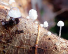
On fallen, decaying leaves of Quercus. Often found under a carpet of leaves and not visible unless the leaves are removed. Rarely found on other substrates, as Quercus fruits, Fagus leaves, and fallen leaves and twigs of Rubus sp. Autumn to late autumn. Widespread in the region but probably often overlooked. In Norway only found in the south east.
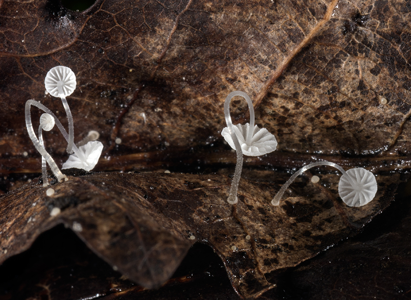
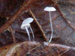
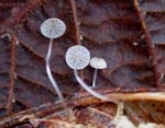
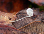
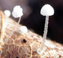
Pileus 1-4 mm across,
covered with a separable, gelatinous pellicle, conical to broadly campanulate, parabolical to convex, sometimes shallowly depressed centrally, sulcate, translucent-striate, pruinose but often appearing glabrous, at
first pale grey or grey-brown, soon becoming more whitish. Lamellae
5-13 reaching the stipe, ascending, adnate, white. Stipe
2-20 mm long, filiform, terete, equal or somewhat wider at the
base, straight to flexuous, glabrous for the greater part,
pruinose-pubescent towards the base, shiny, watery white
to watery grey, sometimes fairly dark grey below; springing from
a small, whitish, pubescent basal disc. Odour
none.
Basidia 13-20 x 6-10 μm, broadly
clavate to obpyriform, 4-spored. Spores
7-12 x 3-4.5 μm, Q 1.8-2.5, Qav ≈ 2, elongated pip-shaped to almost cylindrical,
amyloid. Cheilocystidia
8-30 x 6.5-14 μm, clavate to obpyriform or subglobose,
with fairly few, usually simple, occasionally branched,
curved to flexuous excrescences, 2-18 x 1-2 μm. Pleurocystidia
absent. Lamellar trama dextrinoid. Hyphae
of the pileipellis 1.5-7 μm wide,
diverticulate to smooth, often branched and entwined,
embedded in gelatinous matter, the upper surface of the pileipellis concisting of coarser, diverticulate hyphae 3.5-12.5 μm wide, terminating in clavate to subglobose, diverticulate cells with warts and spiny excrescences 2-10 x0.5-1 μm. Hyphae of the
cortical layer of the stipe 1.5-3 µm wide, smooth. Caulocystidia
9-65(-105) long, usually with an inflated base 3.5-6.5 μm wide, gradually tapering outwards, simple
to branched, flexuous to kinked. Clamp connections present in all tissues.
Mycena mucor is easily identified
on account of the minute size, the pale grey to whitish
pileus, the basal disc, and the occurence on Quercus
leaves. The basal disc is so small that sometimes it can be hard to see, and then it could be mistaken for Mycena polyadelpha, which occurs in the same habitat. M. mucor usually has more developed lamellae, however, and a more "shiny look", and a microscopic investigation will readily reveal its identity. It is a member of Mycena sect. Basipedes, together with
M. rhenana, M.
stylobates , and M. tenuispinosa.
M. rhenana differs in having a nitrous
smell and by occurence on decaying knopper galls on Quercus robur acorn cups or on fallen catkins of Alnus glutinosa, or rarely on fallen debris from Castanea sativa and Corylus avellana. It is characterized by lacking cheilocystidia and by a pileipellis with acanthocysts, and broadly conical caulocystidia with acute, sometimes rostrate to needle-like apex. M. tenuispinosa is poorly known. It differs by having broader
spores and pileal surface densely covered with acute spinules. M. stylobates (which
also can be found on fallen oak leaves) is identified by
the cheilocystidia with coarse, inflated excrescences. Besides,
the margin of the basal disc of M.
stylobates is ciliate, while it is velutinous,
not ciliate, in M. mucor.
Maas Geesteranus (1983 a: 410) described the pileus of Mycena mucor as "greyish or pale grey-brown or greyish beige, whitish towards the margin, pallescent with age". Emmett et al. (2008: 363) described it as greyish brown. In my opinion it rather is white, as shown in the photos at this site.
A somewhat darker species, found on Betula and Salix leaves in alpine areas, was formerly identified as M. mucor but was recently described as a new species and given the name Mycena mucoroides (Aronsen in Aronsen & Læssøe 2016). It differs, besides of having a more brownish cap, in a larger basal disc, broader spores, cheilocystidia with longer and coarser excrescences, a pileipellis with acanthocystoid terminal cells, and much shorter and irregularly shaped caulocystidia. The new species is supported by molecular data.
Microphotographs of the cheilocystidia - 1
Microphotographs of the cheilocystidia - 2
Microphotographs of the cheilocystidia - 3
Microphotographs of the pileipellis- 1
Microphotographs of the pileipellis - 2
Microphotographs of the caulocystidia
Further images on the web:
Mycologie
Normandie |