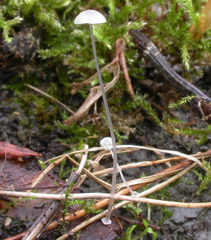
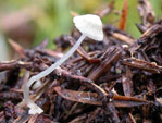
Solitary or in small groups on fallen twigs, leaves, conifer needles,
dead culms of grasses etc.. Summer to autumn. Widespread and fairly common. In Norway found all over the country.
Pileus 4-10
mm across, viscid, covered with a separable, gelatinous
pellicle, conical to planoconvex, flattening with age, translucent-striate,
sulcate, glabrous or occasionally somewhat hispid, pale grey-brown to greyish white. Lamellae
14-18 reaching the stipe, ascending, narrowly adnate to free, often forming
a pseudocollarium, white. Stipe
15-35 x 0.3-1 mm, hollow, fragile, straight, pruinose above, glabrous
farther below, watery grey to white, springing from a basal disc which
is up to 2 mm across, sulcate, pubescent, white, with a
ciliate margin. Odour none.
Basidia 16-25 x 6-8 μm, clavate,
4-spored, with sterigmata up to 5 μm long. Spores 7.5-10.5 x 3.5-5 μm, Q 1.6-2.7, Qav 1.9-2.1, elongated pip-shaped to almost cylindrical,
smooth, amyloid. Cheilocystidia
18-60 x 5-13 μm, forming a sterile band, irregularly clavate, fusiform
or almost cylindrical, with few to numerous variously shaped,
and often coarse excrescences with rounded apices. Pleurocystidia
absent. Lamellar trama dextrinoid. Hyphae of the pileipellis
2.5-6 μm wide, embedded in a gelatinous matter, variously branched, smooth or densely covered with warts or short cylindrical excrescences 1-7 x 0.5-1 μm, terminal cells clavate, up to 11μm wide. Hyphae
of the cortical layer of the stipe 2-4 μm wide, smooth
withscattered caulocystidia
45-80 x 7-8 μm, fusiform, smooth. Clamp connections present in all tissues.
There are several species springing from a
basal disc. Mycena mucor
is quite similar to M. stylobates but is usually smaller and grows on fallen, decaying leaves
of Quercus. The
cheilocystidia are different, with very slender excrescences,
and the margin of the basal disc is not ciliate. M.
bulbosa grows on herbaceous
stalks in wet habitats, the spores are non-amyloid, and
the lamellar edge contains a tough-elastic, gelatinous thread. M. aciculata has a pubescent to setose
pileus (showing as long, thick-walled
cells in the microscope), and an entirely puberulous
stipe springing from a setose basal disc; the
cheilocystidia are different, and the spores are non-amyloid. The closest species is probably M. tenuispinosa. It differs in having a cap densely covered with more prominent spines, cheilocystidia with narrower excrescences, and very conspicuous clusters of pileal spinules springing from the hyphae of the pileipellis.
Microphotos of the hyphae of the pileipellis 1
Microphotos of the hyphae of the pileipellis 2
Microphotos of the cheilocystidia 1
Microphotos of cheilocystidia 2
Next image
|