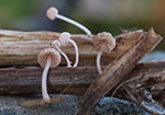
Cespitose or in small groups on decayed
leaf sheaths of Juncus, Typha, Carex and stems
of Scirpus in wet places. Autumn. A rarely recorded species, but known from most countries in the area. Rare in Norway and listed as NT
in the Norwegian red list (2021)
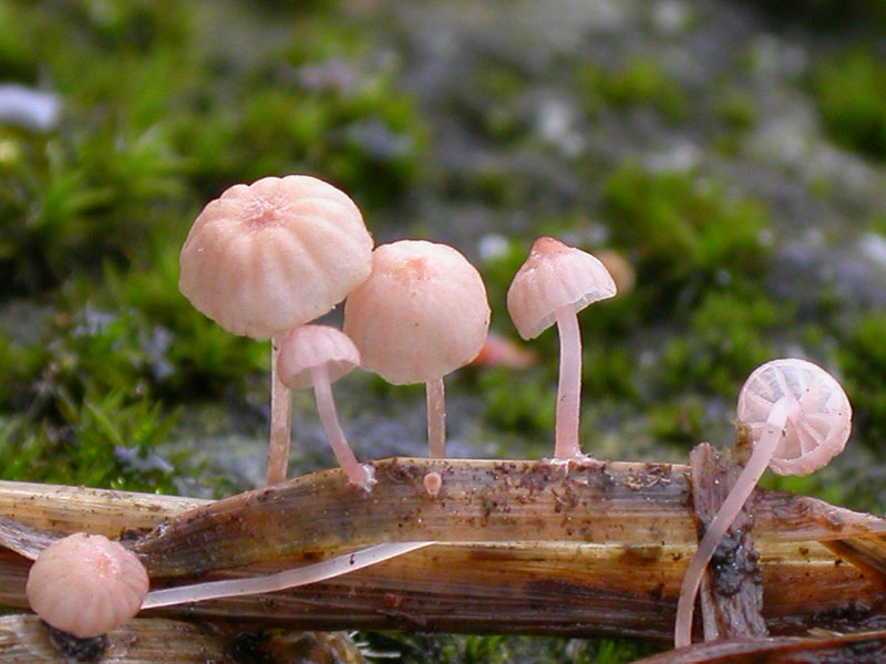
Pileus 2-5(-8) mm across, hemispherical, parabolical, conical, broadly campanulate to almost plane with the margin irregularly
elevated, ± depressed or with a small papilla, margin at first incurved, sulcate, translucent-striate, not hygrophanous, dry, pruinose or finely puberulous, glabrescent, pink to lilaceous pink, darker at the centre, with age brownish beige with no traces of pink. Lamellae 7-11 reaching the stem, very broad, arcuate to subhorizontal,
the edge concave, broadly adnate to subdecurrent, pale pink to whitish; the edge slightly elastic, white-pruinose. Stipe 7-18 x 0.2-0.7 mm, cartilaginous, terete, equal or somewhat wider above (the base sometimes a little thickened), entirely pruinose, glabrescent for the greater part, pale pink or whitish with a pink shade; attached with a whorl of fairly few radiating, white fibrils. Flesh thin, concolorous
with the pileus surface. Odour none. Taste reported as mild (Maire in Kühner 1938).
Basidia 24-32 x 7-12 µm, clavate, 4-spored, with sterigmata 3-6 µm long. Spores (9-)9.8-14 x 4-5(-5.5)
µm, Q 2.3-3.1, Qav ≈ 2.8, elongated pip-shaped, smooth, amyloid. Cheilocystidia
13-30 x 5-13 µm, forming a sterile band, embedded in gelatinous matter
and hard to detect, clavate, covered with rather few to fairly
numerous , more or less evenly spaced, simple, occasionally branched, cylindrical
excrescences 1.5-10 x 1-2 µm. Pleurocystidia absent. Lamellar trama dextrinoid, vinescent in Melzer’s reagent. Hyphae of the pileipellis
2.5-4 µm wide, embedded in gelatinous matter, smooth to densely diverticulate, with simple to branched excrescences 0.5-8 x 0.5-1 µm. Hyphae of the cortical layer of the stipe
3-4.5 µm wide, diverticulate, with simple to branched excrescences 1-6 x 1-2 µm, terminal cells 5-9 µm wide, clavate, diverticulate. Clamp connections present at all tissues.
Microphotos of cheilocystida
Mycena tubarioides occurs in the
same habitats as similar-looking M. culmigena Maas
Geest., M. juncicola, and M. riparia. It differs, however, from these species by the gelatinous
layer covering the pileus, a gelatinous lamellar edge, and larger spores.
Besides, M. riparia
is clampless and has
differently shaped cheilocystidia.
|