On moss-covered trunks of deciduous trees, possibly mostly in rich, calcareous sites. Autumn to early winter. Rare. Listed as near threatened NT in The Norwegian Red List (2021).
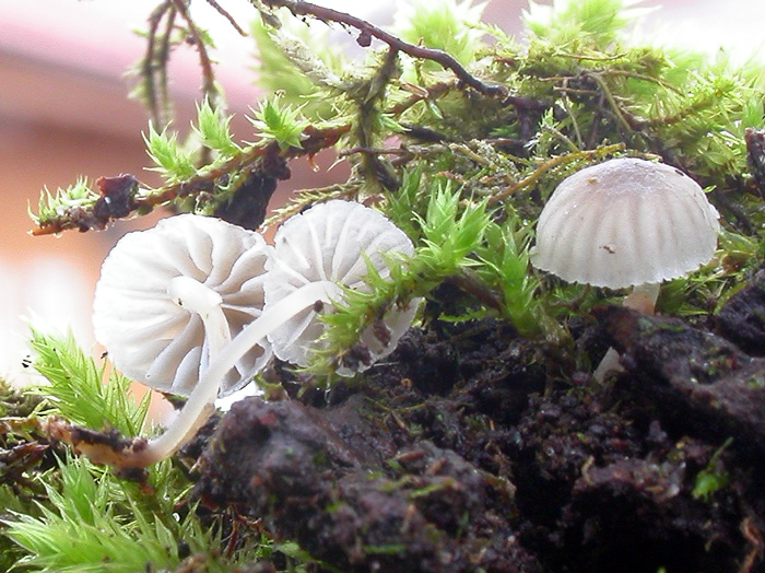
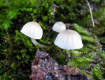
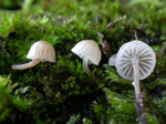
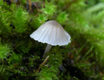
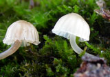
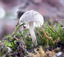
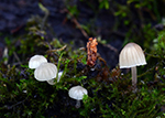
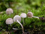
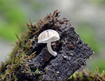
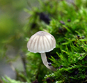
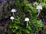
Pileus 2-7 mm across, hemispherical, parabolical to convex, pruinose or minutely puberulous, glabrescent, sulcate, translucent-striate, at first sepia brown with dark sepia brown centre, becoming pale grey-brown more or less with a sepia brown centre and a paler, usually whitish, margin, with age pallescent, turning pale whitish grey to dingy whitish, with or without a darker centre. Lamellae 11-17 reaching the stipe, ascending to subhorizontal, adnate to broadly adnate, decurrent with a tooth,
the edge concave to convex, pale grey to greyish white, edge paler. Stipe 7-15 x 0.5-1 mm,
hollow, terete, equal or somewhat widened at the base, curved to somewhat flexuous, minutely puberulous, glabrescent for the greater part, at first blackish brown, soon becoming paler, pale grey-brown to dingy whitish, often whith a white apex and sepia brown base towards the base densely covered with long, flexuous, white fibrils. Odour none. Taste insignificant, but also experienced as slightly farinaceous.
Basidia 25-35 x 10-12 µm, clavate, 4-spored,
with sterigmata up to 6 µm long. Spores 7-9 x 6.5-9 µm, Q 1.0-1.3, Qav ~ 1.1, globose to subglobose,
smooth, amyloid. Cheilocystidia 15-30 x 8-18.5 µm, occuring mixed with basidia,
clavate, covered with not very numerous, evenly spaced, cylindrical
excrescences 2-5 x 1 µm. Pleurocystidia absent. Lamellar trama dextrinoid, vinescent in Melzer's reagent. Hyphae of the pileipellis 2-7 µm wide, densely covered with cylindrical excrescences 1-2 µm long. Hyphae of the cortical layer of the stipe 1.5-3 µm wide, smooth to sparsely covered with cylindrical excrescences 1-3 x 0.5-1 µm, terminal cells not numerous, 4-6 µm wide, clavate, covered with cylindrical excrescences. Clamp connections present in all tissues.
The description is based on three Norwegian
collections. Mycena supina is rare in Northern parts of Europe
(Maas Geesteranus 1992: 43, Emmett et al. 2008: 382), and the whole variation of this species is probably not yet completely understood. M. supina is a member of sect.
Supinae and occurs in the
same types of habitat as M.
meliigena
and M. pseudocorticola. These both species often turn into brownish colours
with age. They can, however, readily be separated on account
of the cheilocystidia, which are covered with fairly short,
unbranched, cylindrical excrescences in M. supina,
while the cheilocystidia of the two other species are covered
with more or less branched and fairly long excrescences.
In addition the spores of M. supina are smaller
than in M. pseudocorticola and M. meliigena.
Mycena conicoalba Villarreal & Esteve-Rav., described from Spain (Villarreal & Esteve-Raventós 1999), was said to differ from M. supina on account of the persistently conical pileus, the completely white colour of both pileus and stipe except for a yellowish papilla, by the hyphae of the pileipellis which tend to form dense corraloid masses, and the presence of caulocystidia all over the stipe.
In the field Mycena supina can be mistaken for M. alba because of the pale colours and the puberulous pileus. The latter, however, belongs to a completely different section with very different microscopic characters, e.g. smooth cheilocystidia and inamyloid spores. Robich (2003: 670) described the pileus of M. supina as "grigio-bruno, bruno-fuligginoso, bruno-rosso pallido, brunastro, bruno pallido con disco sempre piú scuro". He did not mention the whitish grey to whitish colours of the pileus but claims that a reddish brown colour may be present. It seems to be a question whether his collections of M. supina may have been mixed up with specimens of M. meliigena. His photo of M. supina (Robich 2003: 671) shows much darker specimens than the photos on this site.
Go to key to the
sect. Supinae.
Further images on the web
http://www.naturamediterraneo.com/forum/topic.asp?TOPIC_ID=19500
http://herve.cochard.free.fr/Mycena/Mycena%20supina.htm
|