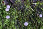
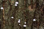
On moss-covered bark of various living deciduous
trees, very rarely on conifer bark (e.g. Abies and Juniperus). Autumn to winter. Widespread and common in most of the covered area, but probably absent in northernmost parts of Scandinavia. Widely distributed and common in South Norway.
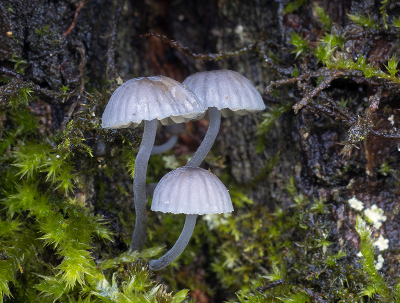
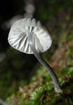
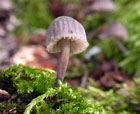
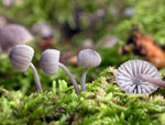
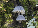
Pileus 2-12
mm across, hemispherical, parabolical, broadly
conical to convex, occasionally with a small papilla, often
somewhat flattened at the centre, sulcate, translucent-striate,
pruinose, glabrescent, dark bluish grey, bluish to bluish
grey, or slate grey, turning brownish with age. Lamellae
8-14 reaching the stipe, fairly broad, ascending to subhorizontal,
the edge convex, broadly adnate, mostly decurrent with a
short tooth, grey to pale bluish grey, or greyish white,
becoming pale sepia brown with age, the edge paler. Stipe
5-25 x 0.2-1 mm, equal, curved, pruinose-floccose, glabrescent, grey
to bluish grey, more brownish with age, the base densely
covered with long, white fibrils. Odour none. Taste insignificant.
Basidia 22-36 x
9-12 µm, clavate, 4-spored or 2-spored, with plump sterigmata up to 13 µm long.
Spores from 4-spored basidia 8-10.5
x 7.5-10 µm, from 2-spored basidia up to 13 x 12
µm, Q 1.0-1.2, Qav~1.1, globose to subglobose, smooth, amyloid. Cheilocystidia
12-54 x 6-25 µm, occuring mixed with basidia,
clavate, covered with unevenly spaced,
simple to branched, curved to tortuous excrescences 0.5-20- x 0.5-1 µm. Pleurocystidia absent. Lamellar trama dextrinoid, staining red-brown in Melzer’s reagent. Hyphae
of the pileipellis 2-7 µm wide,
covered with cylindrical excrescences 0.5-3 x 0.5-1 µm. Hyphae
of the cortical layer of the stipe 2-8 µm wide, diverticulate, with excrescences 0.5-2 x 0.5-1 µm; terminal cells (caulocystidia) 16-37.5 µm long, diverticulate. Clamps present in 4-spored form, absent in 2-spored form.
Mycena pseudocorticola and M.
meliigena can often be
found growing together on the same trunk. M. pseudocorticola
seems to be a little bit more common. Young, fresh specimens
of the two species are not difficult to separate, but with
age they both turn more brownish and can be hard to identify
macroscopically. Microscopically they are very similar too.
Maas Geesteranus (1982a) pointed at a quite reliable character
to tell them apart in the shape and size of the terminal
cells of the stipe cortex. In M. pseudocorticola
these cells are stubby, not longer than 37 µm, whereas
much longer, more slender, cells are by far the more common
kind in M. meliigena.
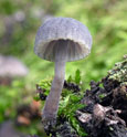
The brown colours in older specimens may cause
confusion with M. supina, but that species have cheilocystidia with
only short excrescences. M.
juniperina has a pale yellowish
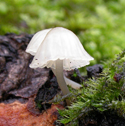
brown pileus and grows on Juniperus communis. Entirely white specimens (with slate grey stipes) of Mycena pseudocorticola may also be encountered (see photo below).
Go to key to sect.
Supinae.
|