|
Generally gregarious on fallen needles of coniferous trees, but
occasionally on litter of deciduous trees. Autumn. Widespread but possibly often overlooked. Widely distributed in Southern Norway but not common.
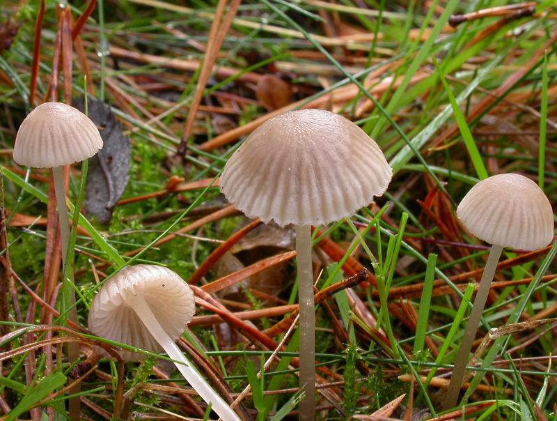
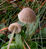
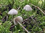
Pileus 5-25
mm across, conical, obtusely conical, campanulate or parabolical,
at age sometimes somewhat depressed centrally, pruinose,
glabrescent, shallowly sulcate, translucent-striate, hygrophanous,
pale grey to greyish brown, somewhat darker (often reddish)
at the centre, the margin often very pale, pallescent when
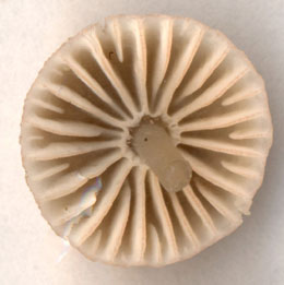
drying. Lamellae
12-20 reaching the stipe, ascending, narrowly adnate,
dorsally intervenose with age, whitish, cream to grey, the
sides densely punctate with minute, dark red-brown dots
(seen under hand lens, but not always easily seen), the
edge dark reddish brown. Stipe
20-70 x 1-1.8 mm, hollow, straight to curved, terete, fragile,
glabrous except for the pruinose apex, becoming shiny, grey,
greyish brown, darker below, the apex whitish to grey; the
base covered with white fibrils. Odour
nitrous.
Basidia 27-30 x 7-9 µm, clavate, 4-spored, with sterigmata 4-6 µm long. Spores
8-11.5 x 4.5-6 µm, Q 1.5-2, Qav ~1.8, pip-shaped to almost cylindrical, smooth, amyloid . Cheilocystidia 35 -
77 x 10 - 16 µm, smooth, fusiform or clavate, apically
generally passing into a simple neck, but also with rounded
apex; with reddish contents. Pleurocystidia
numerous, similar, but sometimes without coloured contents. Lamellar trama dextrinoid.
Hyphae of the pileipellis 2-8 µm wide, covered with simple to branched excrescences 1-30 x 1-3 µm which may form dense masses. Hyphae
of the cortical layer of the stipe 2.5-3.5 µm wide, smooth to sparsely covered with simple, cylindrical excrescences 2-8 x 1-2 µm, terminal cells 4-7 µm wide, coarsely diverticulate. Clamp
connections present in all tissues.
Microphotos of cheilocystidia
Microphotos of cheilocystidia and hyphae of the pileipellis
The nitrous smell is quite strong and reliable
as an identification character. I have not seen the dark
red-brown dots on the sides of the lamellae in all collections.
They can only be seen under a hand lens and even then they
sometimes are hard to discover. The colour of the lamellar
edge is not as dark and distinct as in M.
rubromarginata, but is always visible.
Besides from the smell M.
rubromarginata differs in having more broadly pip-shaped
spores, and usually it grows on decaying wood.
One should also notice a possible confusion
with Mycena olivaceomarginata,
which sometimes has a greyish brown pileus with a reddish
brown centre as well as a stipe with a dingy whitish apex
and greyish brown colours below, and a nitrous smell. The
two species can be told apart on account of the cheilocystidia.
In M. capillaripes they are smooth while they are
more varied in M. olivaceomarginata,
often with two or three necks or with several coarse excrescences.
The latter also lacks pleurocystidia.
Go to Sect. Rubromarginatae.
|