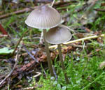
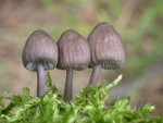
Growing in small groups or subfasciculate
on debris of various deciduous trees or on moss-covered
trunks of living trees. Occasionally also found on coniferous debris (Picea). Occuring typically in early summer,
but can also be found in the autumn. Common in Europe and also widespread in Norway. Not uncommon in Salix scrub in alpine sites.
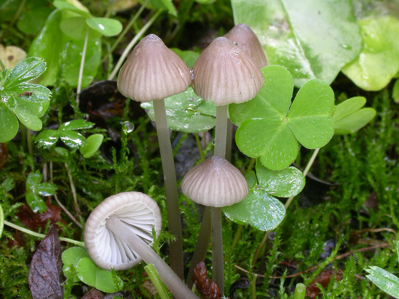
Pileus 10-40
mm across, parabolical to conical, flattening with age,
with or without a low umbo, translucent-striate, sulcate,
pruinose, glabrescent, grey-brown to somewhat darker brown. Lamellae 15-27 reaching the stipe,
ascending, narrowly adnate to adnate, smooth to somewhat
rugulose to strongly veined, becoming dorsally intervenose,
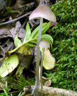
white to greyish with paler edge. Stipe 30-90 x 1-2(-4) mm, fragile, equal or somewhat broadened
downwards, terete, hollow, smooth, pruinose above, glabrous below, becoming shiny when drying, when very fresh exuding a watery fluid
when incised, pale grey, dark grey-brown to dark sepia brown,
usually somewhat darker below and whitish grey at the apex, pallescent with age, the base densely covered with long,
coarse, flexuous, whitish fibrils. Odour more or less nitrous, or sometimes absent. Taste +/- mild.
Basidia 25-36 x
8-9 μm, clavate, 4-spored. Spores 7.5-13 x 4-6.5 μm, Q = 1.6-2.1, Qav = 1.8-1.9, elongated to cylindrical, smooth, amyloid. Cheilocystidia 25-55 x 10-14 μm, fusiform, clavate, subcylindrical,
more rarely sublageniform, usually apically drawn into a
slender neck or occasionally with two or more necks. Pleurocystidia similar. Lamellar trama dextrinoid, brownish vinescent in Melzer's reagent. Hyphae of the pileipellis 2-4 μm wide, covered with scattered to crowded, simple to branched, excrescences 2-10 x 1-2 μm, which tend to become gelatinized. Hyphae of the cortical layer of the stipe 2-2.5 μm wide, sparsely diverticulate, terminal cells hard to find, uniflated, diverticulate. Clamp connections present in all tissues.
Microphotos of cheilocystidia
Microphotos of the hypae of the pileipellis
Microphotos of spores
Mycena abramsii is a member of the
large section Fragilipedes, where it takes a position close to section Lactipedes. The watery exudation produced by incising
the stipe is a distinct feature of this species, but is
rarely demonstrable in specimens only a few hours after
collecting, and in fresh specimens that have become somewhat
dry.
It is not always easily distinguished from
other members of the section. The cheilocystidia, which
apically usually are drawn into a slender neck, and the
somewhat elongated spores are useful identification characters.
The smell is variable and has been recorded differently
by different collectors, but typically is rather faint.
M. aetites differs in having shorter but broader
spores and differently shaped cheilocystidia, as well as
in the habitat; lawns, meadows and grassland. In M.
leptocephala the hyphae of the cortical
layer of the stipe are smooth, the terminal cells are generally
conspicuously inflated, and the cheilocystidia are somewhat
differently shaped. M. galericulata, which occurs in the same habitats, is tough (especially the stipe), and the cheilocystidia are densely covered with simple to branched, finger-like excrescences.
|
_small.jpg)