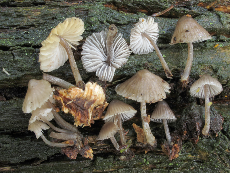
Single or in small groups on wood of Quercus, often occuring with M. galericulata and M. inclinata. Autumn. Known from a few sites in old-growht woodland in Germany, Great Britain and Denmark (Læssøe 2013 b), but not yet in Norway.
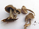
Pileus 4-25 mm across, as young parabolic, then campanulate with a small umbo, in young specimens with a crenulate margin (as in M. inclinata), hygrophanous, sulcate, translucent-striate, at the centre and the margin "vernissiert-bereift" (as in M. filopes) and dry, or not pruinose and somewhat viscid, dark grey-brown, fading to pale grey-brown when drying, often reddening with age. Lamellae 15-22 reaching the stipe, ascending, narrowly adnate, decurrent with a small tooth, becoming dorsally intervenose with age, grey, the edge concolorous. Stipe 13-55 x 1-2.5 mm, hollow, somewhat widened towards the base, conspicuously white-pruinose, glabrescent except for the pruinose apex, grey to grey-brown, dry, somewhat shiny, the base covered with white, flexuouse fibrils. Odour indistinctive to weakly raphanoid. Taste indistinctive to somewhat disagreeable, of cucumber.
Basidia 30-39 x 7-10 µm, clavate, 4-spored, with sterigmata up to 10 µm long. Spores 7-10 x 6-7.5 µm, Q 1.2-1.6, Qav≈1.3, broadly pip-shaped, smooth, amyloid. Cheilocystidia 42-74 x 18-40 µm, occuring mixed with basidia, fusiform to lageniform or clavate to broadly clavate, sometimes spheropedunculate, with slightly thickened cell walls in the widest part, apically densely covered with simple, cylindrical excrescences up to 3 µm long (hardly visible in dried material). Pleurocystidia numerous, similar. Lamellar trama dextrinoid. Hyphae of the pileipellis 2-3 µm wide, smooth to sparsely covered with thin excrescences 0.5-2 x 0.5-1 µm. Hyphae of the cortical layer of the stipe 1.5-5 µm wide, smooth to sparsely covered with straight to curved excrescences 1-7 x 0.5-2 µm, caulocystidia numerous, 23-110 x 5-25 µm, subcylindrical to clavate, covered with few to fairly numerous, rather coarse excrescences. Clamp connections present in all tissues.
The macroscopic description has been taken from the type description, complemented by observations on a Danish collection. The microscopic details are based on examination of one Danish and one German collection.
Next image
Microphotos of cheilocystidia 1
Microphotos of cheilocystidia 2
Microphoto of a caulocystidium
Mycena silvae-pristinae belongs to sect. Intermediae on account of the large cheilocystidia and strongly protruding pleurocystidia, apically covered with excrescences. It is closely related to the American species Mycena borealis A.H. Smith, from which it differs in smaller size, dry to somewhat viscid pileus, conspicuous caulocystidia, and the excrescences on the hymenial cystidia disappearing in exsiccates. In addition M. borealis grows on coniferous wood. In the field M. silvae-pristinae may resemble M. inclinata that is found in the same habitat, but it differs entirely in the microscope.
The phenomenon that the excrescences at the hymenial cystidia disappear in dried material, was also mentioned by Veerkamp & Kuyper (1997). The excrescences are very visible in fresh material, while in dried material they become covered by a glutinous matter and are hardly visible. Typically, masses of spores are then sticking to the apex of the cheilocystidia. |