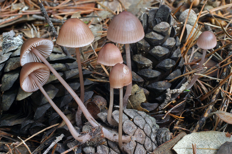
Solitary to subfasciculate on fallen Pinus cones, occasionally also on moss-covered, decaying wood of a Pinus trunk; mainly but not exclusively found in coastal areas. Autumn. Occuring north to France. Not in the Nordic countries.
Pileus 25-40 mm across, parabolical or conical to campanulate, finally more or less flattening, not or very shallowly sulcate, translucent-striate, innately fibrillose to polished, dry but slightly lubricous when moist, pale purplish brown to brownish beige with chestnut brown to dark purplish brown centre, fading to dingy flesh-colour wiyh vinaceous and yellowish tints, the margin pink or lilaceous to fairly dark vinaceous. Lamellae 18-23 reaching the stipe, ascending, adnate to somewhat decurrent with a short tooth, with age dorsally intervenose, dingy whitish, pale greyish to brownish, becoming flushed with pink or lilac, the edge purplish to dark red-brown. Stipe 40-70 x 1.5-7 mm, hollow, fragile, equal or gradually widening below, terete to laterally compressed, straight to flexuous, curved below, innately fibrillose, glabrous, occasionally sparsely and minutely floccose above, shiny, lilaceous to vinaceous above, gradually turning more brownish, watery white to dingy whitish below, the base passing into a shorter or longer root, densely covered with long, coarse, flexuous, whitish fibrils. Odour faint, agreeable. Taste indistinctive or said to be somewhat raphanoid.
Basidia 29.5-39 x 8-12 µm, clavate, 4-spored, with sterigmata up to 6 µm long. Spores 10.3-14 x 6-7 µm, Q 1.6-2.2, Qav≈1.9, pip-shaped to somewhat elongated, occasionally somewhat constricted in the middle part, smooth, amyloid. Cheilocystidia 28-47 x 7.5-14 µm, forming a sterile band, fusiform, subcylindrical, lageniform to clavate, smooth, occasionally with one or more, coarse excrescences apically, with reddish brownish contents. Pleurocystidia scarse, occuring near the lamellar edge, similar. Lamellar trama dextrinoid. Hyphae of the pileipellis 1.5-5 µm wide, with a tendency to become gelatinized, with brownish contents, smooth or sparsely covered with rather coarse excrescences 2-5 x 2-3.5 µm. Hyphae of the cortical layer of the stipe 1-5 µm wide, somewhat gelatinized, smooth, terminal cells 3.5-9 µm wide, smooth to coarsely diverticulate. Clamp connections present in all tissues.
The macroscopic description has been taken from Maas Geesteranus(1986). The microscopic details are based on examination of one French collection.
Mycena seynii is a member of sect. Rubromarginatae, where it takes place close to M. rubromarginata. It differs from the latter on account of the hyphae of the pileipellis, which are smooth or covered with scattered, simple, fairly coarse excrescences (simple to very much branched, forming dense masses in M. rubromarginata), narrower spores, and different terminal cells (more branched in M. rubromarginata), in addition to the different habitat. It should be easy torecognize in the field when it is found growing on Pinus cones, but it should be noted that M. rubromarginata at least can grow on Larix and Picea cones.
|