On decayed stumps of Fagus. One record in Norway.
Known also from Italy, Spain, France, the Czech Republic, Poland, and Austria.
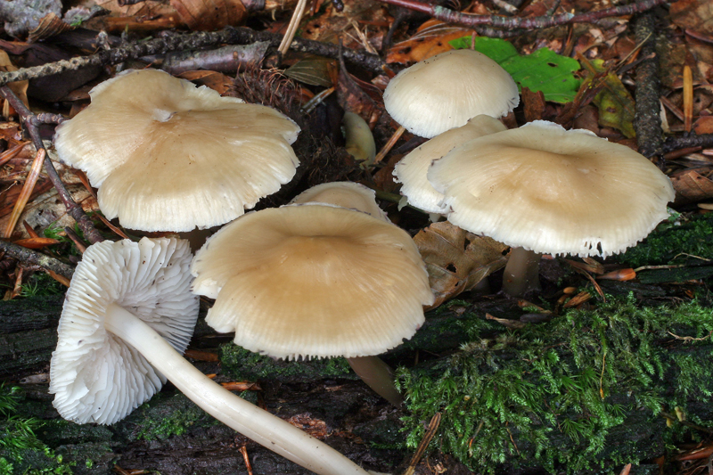
Pileus 20-50 mm across, conical to campanulate, more or less with an obtuse umbo, flattening with age, rugulose at the margin, becoming entirely rugose-sulcate, apparently not translucent-striate, glabrous, not pruinose, young pale greyish brownish to pale orange brownish, darker at the centre, paler nearer the margin, becoming paler with age, pale cream to very pale flesh-colour, whitish at the very margin. Lamellae 27-30 reaching the stipe, elastic-tough, ascending, narrowly adnate, with or without a short decurrent tooth, creamy white, the edge white. Stipe 40-110 x 2.5-6 mm, hollow, tough, equal for the reater part, terete, somewhat curved, smooth, glabrous, not visibly pruinose at the apex, shiny, cream to whitish cream, the base white-tomentose, extending into a long root penetrating the substrate. Odour of apples when cut, then sweetish fruity. Taste indistinctive, not farinaceous.
Basidia 28-38 x 7.5-8 μm, clavate, 4-spored, with sterigmata up to 8 μm long. Spores 8-10 x 5.5-6.5 (-7.5) μm, Q 1.4-1.6; Qav~1.5, broadly pip-shaped to subglobose, smooth, amyloid. Cheilocystidia 24-44 x 7-14.5 μm, forming a sterile band, fusiform to clavate, sometimes with new heads apically, covered with rather few, unevenly spaced, cylindrical, simple, more or less curved excrescences 1.5-7 x 1-2 μm. Pleurocystidia absent. Lamellar trama dextrinoid, vinaceous in Melzer's reagent. Hyphae of the pileipellis 2-5 μm wide, smooth or sparsely covered with low warts or short, cylindrical excrescences up to 2 x 1 μm, terminal cells up to 6.5 μm wide, covered with more prominent excrescences 1.5-9 x 1-3.5 μm. Hyphae of the cortical layer of the stipe 1.5-3 μm wide, smooth or very sparsely diverticulate. Clamp connections present in all tissues.
Microphotos of cheilocystidia
The macroscopic description has been taken from Maas Geesteranus (1991) and Komorowska (2010). The microscopic detailes are based on examination of a French collection kindly put to my disposal by Mr. H. Cochard.
Mycena romagnesiana could easily be mistaken for the more variable M. galericulata. Apart from the different smell and taste, it can be told from M. galericulata by the pileus lacking a translucent striation, shorter spores, the hyphae of the pileipellis and stipitipellis being smooth or only sparsely covered with short excrescences, and the cheilocystidia having fewer and shorter excrescences.
Further images on the web:
Josef Hlasek |