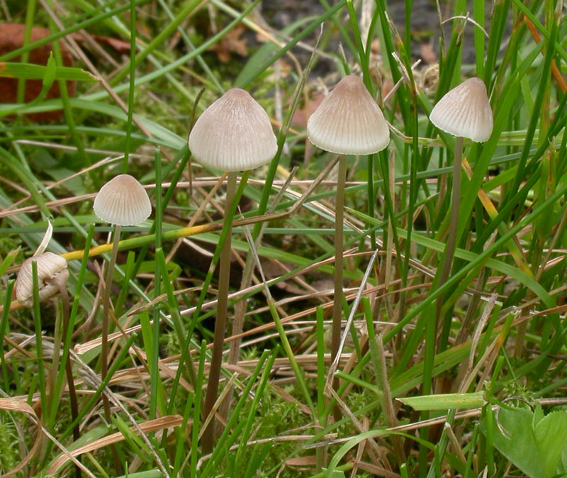
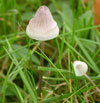
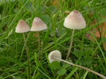
Solitary or in small groups in mossy lawns,
under conifers as well as deciduous trees, on vegetable
debris, decaying wood, not infrequently also on moss-covered
tree trunks. Summer to autumn. Common. Widely distributed in Norway.
Pileus 5-20
mm across, conical to campanulate, often shallowly umbonate,
translucent-striate, shallowly sulcate, pruinose, glabrescent,
finely innate-fibrillose, with the fibrils gradually splitting,
giving the surface a rimose aspect much in the manner of
some species of Inocybe, and imparting a silvery
lustre to the pileus when drying out, hygrophanous, very
pale to almost white, greyish, grey-brown to very dark brown,
at the centre darker, paler to almost whitish towards the
margin. Lamellae 16-23 reaching
the stipe, ascending, narrowly adnate, rarely decurrent
with a small tooth, whitish to pale grey or with a brownish
tinge, as a rule not turning pinkish. Stipe
40-155 x 0.5-2 mm, hollow, equal, straight
or somewhat curved, terete, fairly firm, at first entirely
pruinose or minutely puberulous, glabrescent for the greater
part, pale to fairly dark grey-brown, paler to almost white
at the apex, the base covered with long, coarse, flexuous,
whitish fibrils. Odour indistinctive
when fresh, of iodoform on drying out. Taste mild.
Basidia 20-28 x
8-12 µm, clavate, mostly 2-spored, but also less frequently
4-spored. Spores 9-11.5
x 5.5-6.5 µm (from 2-spored basidia), Q 1.6-1.8, Qav~1.7, or 8-9 x
5.5-6.5 µm (from 4-spored basidia), pip-shaped, smooth, amyloid.
Cheilocystidia
12-30 x 7-18 µm, forming a sterile band, sessile
(often the majority) to stipitate, ellipsoid, obovoid, obpyriform,
clavate, almost spheropedunculate to somewhat irregularly
shaped, covered with fairly few simple to somewhat
branched cylindrical excrescences 0.5-20 x 0.5-1.5 µm. Pleurocystidia absent
or rare, similar. Hyphae
of the pileipellis 1.5-4 µm wide,
densely covered with warts or short cylindrical excrescences 0.5-3 x 0.5 µm, more or less forming dense, corraloid masses. Hyphae
of the cortical layer of the stipe 2-4 µm wide,diverticulate, excrescences 0.5-3.5 x 0.5 µm. Caulocystidia
(terminal cells), abundant in the apical part of the stipe, 5-12 µm wide, clavate to subglobose, more or less curved outwards,
covered with warts. Clamp connections present in all tissues both in the 2-spored as well as in the 4-spored form.
Mycena filopes is one of several greyish to grey brown Mycenas with no good characters to separate them from other similar looking species. It can be particularly difficult distinguish from M. metata. They can be told apart as follows
(taken from Maas Geesteranus 1992):
Mycena filopes:
a) Pileus surface giving the impression of being rimose,
with the innate fibrils splitting much in the way of the
superficial fibrils of the pileus of some Inocybe, and b)
imparting a silvery lustre on drying out, c) pileus not
becoming tinged with pink, d) lamellae not or only rarely
turning slightly pinkish, e) sessile cheilocystidia often
more frequent than the stipitate ones, f) stipitate cheilocystidia
up to 30 µm long, g) terminal cells of the stipe cortex
always present, numerous and easy to find.
Mycena metata: a)
Pileus surface either not rimose or without apparent texture,
b) without silvery lustre, c) pileus and / or lamellae usually
becoming tinged with pink, d) stipitate cheilocystidia often
more frequent than the sessile ones, e) stipitate cheilocystidia
often greatly varying in size and the bigger ones usually
more voluminous than their counterparts in M. filopes, reaching
more than 70 µm in length, f) terminal cells of the
stipe cortex absent or, if present, rare and mostly hard
to find.
The smell of iodoform is best experienced
when the fungus has been kept in a closed box for a while.
Another species that could be mistaken for M. filopes in the fiels is M. vitilis. The two species can occur in the same habitats and sometimes they look quite similar. The latter, however, has completely different cheilocystidia and a more cartilaginous stipe.
Microphotos of the cheilocystidia.
Go to key to
sect. Filipedes.
Further images on the Internet:
Kulakbiocampus/paddestoelen |