|
Mycena simia Kühner
|
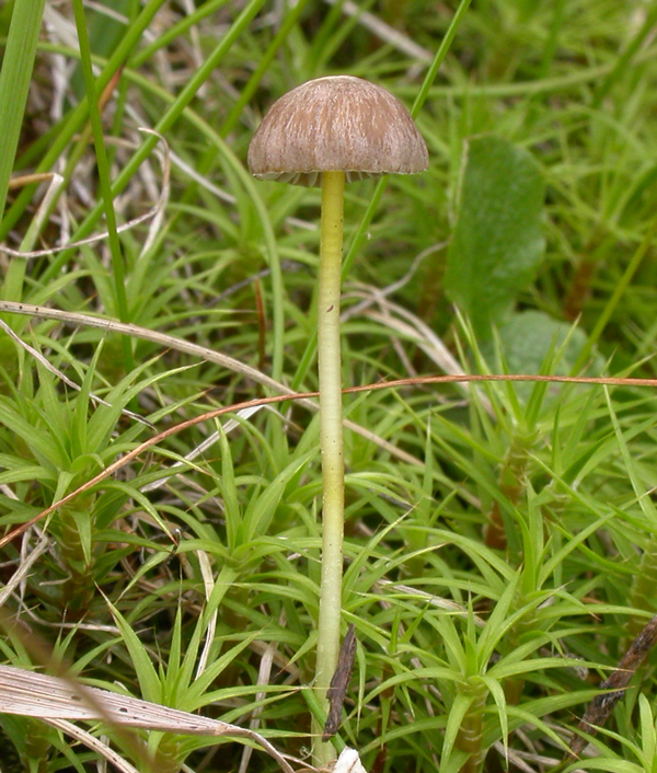
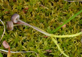
Solitary to gregarious in damp moss including Sphagnum, Polytrichum, Aulacomnium and Hylocomium, in mires and dwarf shrub heaths, along brooks and roadsides, often on north slopes on acid soil. Common in arctic and subalpine areas. Summer to autumn. Pileus 8-13 mm across, conical to campanulate, translucent-striate, sulcate, olive-brown, dark sepia brown to greyish black, sometimes with honey yellow to whitish margin, hygrophanous, covered with a separable, tough, gelatinous pellicle. Odour and taste pronounced, farinaceous-acid. Gills 16-22 reaching the stipe, ascendant, narrowly adnate, decurrent with a tooth, edge gelatinized, separable as a tough, elastic thread, white, pale to dark grey with paler edge. Stipe 20-100 x 1-2.5 mm, viscid, citrine yellow at first, turning dark greyish brown above with age, retaining the yellow colour in lower parts. This taxon is not unusual to find in alpine and subalpine areas in Norway. (See Gulden & Jenssen, 1982). The dark pileus, which can be almost black at the center, the 2-spored basidia, and the absence of clamp connections are important characters. I have occasionally seen a few 4-spored basidia on the same lamella that is dominated by the 2-spored basidia. Phylogenetic analyses of ITS sequences obtained by the Norwegian Barcode Project (NorBOL) are showing that M. simia is a good species clearly separated from M. epipterygia.
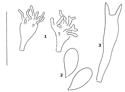
Fig.1. - 1. Cheilocystidia. 2. Spores. 3. Basidium. Bar = 20 μm. Lange (1955) described M. epipterygia var. badiceps from South Strømfjord in Greenland. The variety was described with the cap as “bistre to almost black in centre” and a lemon yellow stipe that becomes dark greyish brown on upper part with age, a pronounced farinaceous-acid smell, 2-spored basidia and large spores (9-12 x 5.5-7.3 µm). He claimed to have seen clamps in the hyphae of the pileipellis but Maas Geesteranus (1989) who reexamined the holotype emphasized that it is clampless (although cheilocystidia occasionally were seen with an abortive clamp). Maas Geesteranus (1980) accepted M. simia as a good species with reference to the fact that Lamoure (Kühner & Lamoure 1958) had shown that M. simia was cytologically different from 4-spored forms of Mycena epipterygia, and from this concluded that both are specifically different. At the time he treated the equally 2-spored M. epipterygia var. badiceps as a separate entity but mentioned that M. simia and M. epipterygia var. badiceps appear very close and “further research may prove them identical”. Later, Maas Geesteranus (1989) reduced M. simia to be a synonym to M. epipterygia var. badiceps, although still with a question mark. When describing M. simia, Kühner (1958) did not mention the similarity to M. epipterygia var. badiceps, which had been described three years earlier. The descriptions, however, look very similar and without having seen the type we feel confident that they represent the same taxon. We have observed a great variability both in the size and shape of the spores in the collections that we have examined, and the different colouring of the stipe is probably not enough to justify two different taxa.
|