Generally associated with conifers, Picea,
Pinus, Juniperus, but occasionally found
in deciduous woods. Widespread in area but local and rare in western lowlands. Widely distributed in South Norway,
although not common. Recorded north to Troms county. Summer to late autumn.
Pileus 10-25
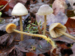
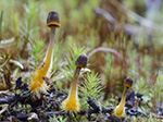
mm across, conical to parabolical, flattening with age, becoming convex to plane, with or without umbo, shallowly sulcate, translucent-striate, glabrous, hygrophanous, grey-brown, olivaceous brown to fairly dark brown with
the margin orange to orange-yellow. Lamellae 16-26
reaching the stipe, ascending, narrowly adnate, decurrent
with a short tooth, pale yellowish grey to greyish beige with bright orange to pale orange-yellow edge. Stipe
35-80 x 1-2 mm, hollow, straight to curved, terete, equal,
firm, entirely pruinose when young, glabrescent except for the apex, whitish to yellowish at the apex, yellow brown
to dark brown farther down or also orange yellow near the base, sometimes tinged with yellowish or orange shades; the base densely covered with yellow
to orange fibrils. Odour very
conspicuous; sweet, fruity, often experienced as farinaceous or faintly of anise.
Basidia 24-33 x 5.5-8 µm, slender-clavate, 4-spored, with sterigmata 4-6.5 µm long. Spores 7.5-10.5 x 4-5.5 µm, Q 1.7-2.1, Qav 1.9, elongated pip-shaped to subcylindrical, smooth, amyloid. Cheilocystidia 18-42 x 7-17 µm, forming a sterile band, variable shape but often clavate to obpyriform, long- to short-stalked, with orange contents, covered with fairly numerous, evenly spaced, simple, cylindrical excrescences 1-3 x 0.5-1 µm. Pleurocystidia similar. Lamellar trama dextrinoid. Hyphae of the pileipellis 1.5-5.5 µm wide, densely covered with simple to branched excrescences 1-24 x 1-3 µm, forming dense masses and tending to become somewhat gelatinized. Hyphae of the cortical layer of the stipe 1.5-3.5 µm wide, smooth to sparsely diverticulate, excrescences simple to furcate 1-9 x 1-1.3 µm, terminal cells 4.5-10 µm wide, simple to variously branched, subcylindrical to clavate, almost smooth to densely diverticulate. Clamp connections present in all tissues.
Microphotos of cheilocystidia.
Microphotos of pileipellis and terminal cells of stipitipellis.
Mycena aurantiomarginata is readily identified on account of the orange lamellar edge and the yellow to orange fibrils at the base of the stipe. It is a member of sect. Luculentae Maas Geest.
 Next image Next image  Next image2 Next image2
|