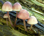
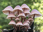
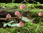
In small groups or fasciculate on fallen
branches and decayed wood of deciduous trees, less frequently
on conifers. Summer to autumn. Common and widespread in the area covered.
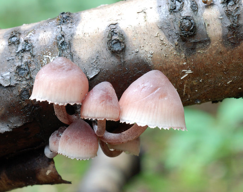
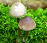
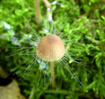
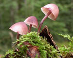
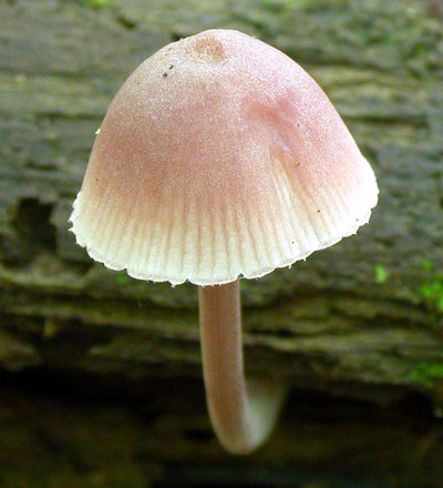
Pileus up to 25(-40)
mm across, obtusely conical, campanulate to parabolical, somewhat flattening
with age, with or without umbo, shallowly sulcate, translucent-striate,
densely powdered or pruinose, glabrescent, hygrophanous,
dark brown with a faint vinaceous shade at the centre, paler
brown with a pinkish shade towards the margin, sometimes
stained with purplish spots with age, drying pinkish white,
the margin usually crenate. Lamellae
18-25 reaching the stipe, ascending, narrowly to broadly adnate, decurrent with a tooth, becoming dorsally intervenose, whitish to grey, developing more brownish flesh-colour or pale vinaceous, sometimes becoming stained with purplish spots in age; the edge
concolorous, sometimes purplish brown to blackish brown.
Stipe up to 60 x 2.5 mm, hollow,
fragile, straight to curved, equal, terete to compressed,
at first densely white-powdered, soon glabrescent, vinaceous
red-brown, darker below, exuding a dark red-brown fluid
when cut; the base densely covered with white fibrils. Odour
indistinctive.
Basidia 27-37 x 8-11 µm, clavate, 4-spored, with sterigmata 5-7 µm long. Spores 8-9.5 x 5-6.5 µm, Q 1.2-1.5, Qav = 1.4, rather broadly pip-shaped, smooth, amyloid. Cheilocystidia 36-70 x 9-15 µm, forming a sterile band, fusiform, with or without reddish brown contents, apically passing into a usually slender neck, more rarely with the neck somewhat branched. Pleurocystidia similar, if present. Lamellar trama dextrinoid, vinescent in melzer's reagent. Hyphae of the pileipellis 2-4.5 µm wide, covered with fairly coarse, diverticulate excrescences 1-12 x 1-3 µm. Hyphae of the cortical layer of the stipe 2-3.5 µm wide, smooth to sparsely covered with short cylindrical excrescences near the terminal cells; caulocystidia 20-55 x 3.5-12.5 µm, occuring in clusters, clavate to irregularly shaped, branched to very coarsely diverticulate. Clamp connections abundant at all tissues.
Microphotos of cheilocystidia 1
Microphotos of cheilocystidia 2
Microphotos of the hyphae of the pileipellis
Microphoto of the hyphae of the cortical layer of the stipe
Microphotos of terminal cells (caulocystidia)
Microphotos of terminal cells (caulocystidia) -2
Lange (1914) described M. haematopus var. marginata, characterized by the lamellae having a reddish edge. Several authors, however, (e.g. Maas Geesteranus 1988d) have stated that the colouration of the lamellar edge in M. haematopus is too variable to have any taxonomic significance.
M. haematopus is the only member of sect. Galactopoda (Earle) Maas Geest., but it is not always easy to tell it apart from M. sanguinolenta belonging to sect. Sanguinolentae Maas Geest. The cheilocystidia of M. sanguinolenta are generally more sharply acuminate, but an even better feature to differentiate the two species is the shape of the caulocystidia. In M. sanguinolenta they are usually narrowly tipped.
|