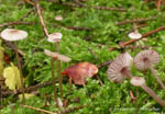
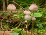
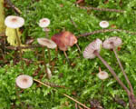
Gregarious on humus and vegetable debris among grass
and moss, on fallen twigs and moss-covered trunks of both decidious
trees and coniferous trees, and among fallen needles of coniferous trees. Early summer to late autumn. Very common and widespread in the covered region, but apparently declining and red listed in the Netherlands (Aronsen & Læssøe 2016). Widely distributed and one of the most common Mycena-species in Norway.
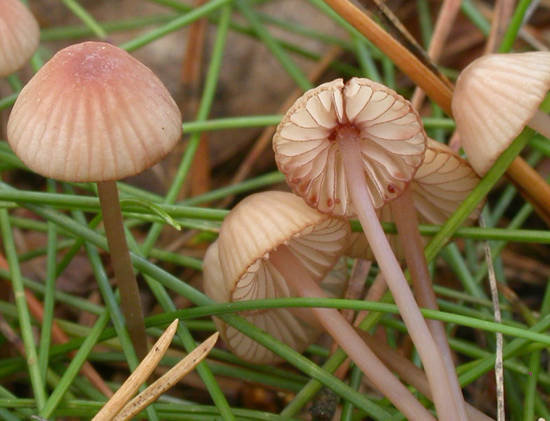
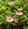
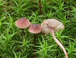
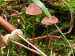
Pileus 5-18
mm across conical, campanulate, convex, flattening to applanate
with age, sometimes with recurved margin, often papillate
or umbonate, but even sometimes somewhat depressed centrally,
sulcate, translucent-striate, very pale brown to grey brown
with dark reddish brown centre, dark brown to pinkish beige
at the centre, pale brown to brown-violet towards the margin.
Lamellae 13-17
reaching the stipe, ascending, narrowly adnate, decurrent
with a short tooth, white or greyish, the edge vinaceous
red or red brown. Stipe 30-60 x
0.5-1.5 mm, hollow, terete, straight to curved, equal or somewhat
widened below, minutely puberulous at the apex, glabrous
farther down, beige brown to brown with a vinaceous or violet
tinge , the base densely covered with white fibrils. With
red to sometimes colourless fluid. Odour indistinctive. Taste mild.
Basidia 27-35 x 8-10 µm, clavate, 4-spored. Spores 8-10(-12) x 4-6 µm, Q 1.6-2.4, Qav ~ 1.9, pip-shaped, smooth, amyloid. Cheilocystidia 27-55 x 6.5-10 µm, forming a sterile band, fusiform, smooth, with reddish brown contents, apically narrowed into an acute neck, but occasionally with two necks or with coarse, lateral excrescences. Pleurocystidia similar, if present. Lamellar trama dextrinoid. Hyphae of the pileipellis 2-4.5 µm wide, covered with simple to somewhat branched excrescences 2-15 x 1.5-2 µm, which may form dense masses. Hyphae of the cortical layer of the stipe 1-3.5 µm wide, sparsely covered with simple to furcated, cylindrical excrescences 1.5-10 x 1-2 µm, caulocystidia 18-55 x 5.5-9 µm, resembling the cheilocystidia but occasionally also diverticulate; at the apex sometimes also some "hair-like" cells. Clamp connections present in all tissues.
Microphotos of cheilocystidia
Microphotos of the cortical layer of the stipe
Microphotos of caulocystidia
This species can be identified on account of the pink to vinaceous shades at the pileus, white to grey lamellae with red brown edge and a red brown fluid both in the pileus and the stipe. When incised the fluid is visible especially at the lamellae and the margin of the pileus. When working with laboratory material and dry specimens, the sharply acuminate cheilo- and caulocystidia are useful characters.
Dry specimens (when the fluid is not visible) can be confused with M. rubromarginata. They have the red gill edge in common, but the latter has broader spores and much longer excrescences on the pileipellis hyphae, and the cystidia are not sharply acuminate.
Further images:
|