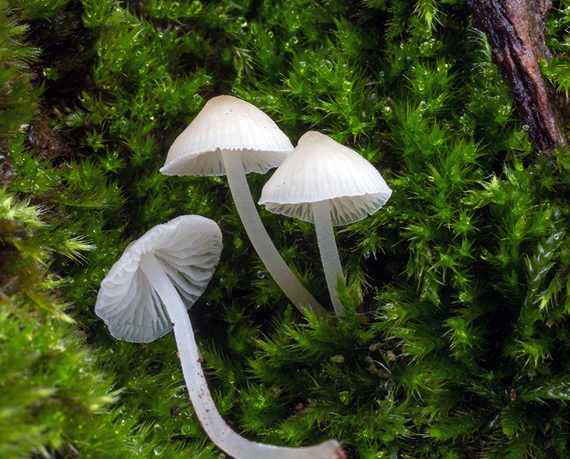
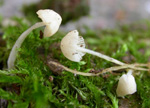
Solitary to scattered or in small groups
on moss-covered trunks of deciduous trees. Autumn. Not common. Listed
as NT in The Norwegian redlist.
Pileus 5-15
mm across, conical or somewhat campanulate to convex, flattening
with age, occasionally with a small papilla, glabrous, hygrophanous,
translucent-striate, sulcate or not, whitish to ivory, becoming
more yellowish with age, especially in the centre. Lamellae
15-22 reaching the stipe, ascending, narrowly adnate to
emarginate, sometimes decurrent with a short tooth, somewhat
veined with age, and then often intervenose, anastomosing,
white. Stipe 15-30 x 0.5-2 mm,
straight to curved, equal, terete, firm, minutely puberulous
all over, glabrescent, becoming shiny, white, turning yellowish
to yellow-brown from the base with age; the base densely
covered with long, coarse white fibrils. Odour
not distinct. Taste mild or faintly acidulous.
Basidia 22-27 x 6-7 µm, clavate,
2-spored, not clamped, with sterigmata up to 8 µm long. Spores
6.5-9.5 x 4.7-6.2 µm, q = 1.3 - 1.8, qav ~ 1.5, broadly pip-shaped, smooth,
nonamyloid. Cheilocystidia
35-60 x 10-20 µm, occuring mixed with basidia, cylindrical, subclavate, fusiform,
apically broadly rounded or somewhat narrowed, smooth, clampless.
Pleurocystidia scattered, similar. Lamellar trama not vinescent in
Melzer's reagent. Hyphae
of the pileipellis 2.5-4.5 µm wide, smooth, sometimes with a few scattered, coarse, cylindrical excrescences, clampless, terminal cells 4-8 µm wide, sometimes with a few coarse, inflated excrescences. Hyphae of the cortical layer of the stipe
2-4.5 µm wide, smooth, clampless. Caulocystidia
16-48 x 5-9 µm, simple or lobed to somewhat branched, clavate or fusiform to more
irregularly shaped.
P minutula may show similarity to P.
flavoalba, but that species
does not grow on wood; it has differently shaped cheilocystidia
and caulocystidia, the spores are narrower, and the hyphae
of the pileipellis are diverticulate. In P.
hiemalis the hyphae
of the pileipellis are diverticulate, and the cheilocystidia
are more fusiform or utriform. P.
alba, also frequenting the same habitats,
can be distinguished on account of the arcuate to horizontal,
broadly adnate to somewhat decurrent lamellae, the globose
spores and the inflated excrescences of the hyphae of the
pileipellis.
The odour of Phloeomana minutula has been variably described by different authors. In the Latin protologue Bresadola described it as "odore forti rancido praedita" (Bresadola 1881: 73). Kühner (1938: 568) described the odour as "faible (un peu fruitée ou spermatique)". Maas Geesteranus (1991: 86) described it as "none, faint or hard to describe". Robich (2003: 371), however, claimed that it is indistinctive or weakly raphanoid. According to my experience the odour is not distinctive.
According to Kühner (1938) P. minutula can also occur with 4-spored basidia. This was repeated by Emmett et al. (2008). All the collections examined by me, however, have been 2-spored.
The variation of both P. minutula and P. hiemalis does that separating the two species is not always easy. Specimens of P. hiemalis occasionally are entirely white, and the pileus of P. minutula often shows a yellowish centre. Besides, the hyphae of the pileipellis in P. minutula are not always entirely smooth. The photo in Robich (2003: 369) shows specimens with a very dark centre, resembling P. hiemalis indeed, and the photo of P. hiemalis shows specimens with a yellowish white pileus, indicating that the two photos must have been mixed up.
Microscopic drawings
More microscopic drawings
Further images on the web:
|