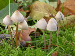
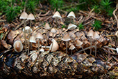
In groups in mossy lawns, under both conifers
(mostly Picea) and deciduous trees, on fallen twigs
and other vegetable debrise, not infrequently also on moss-covered los. Often very numerous on needle
beds. Summer to late autumn. Very common. Widely distributed. Also in alpine sites. Recorded in all parts of Norway.
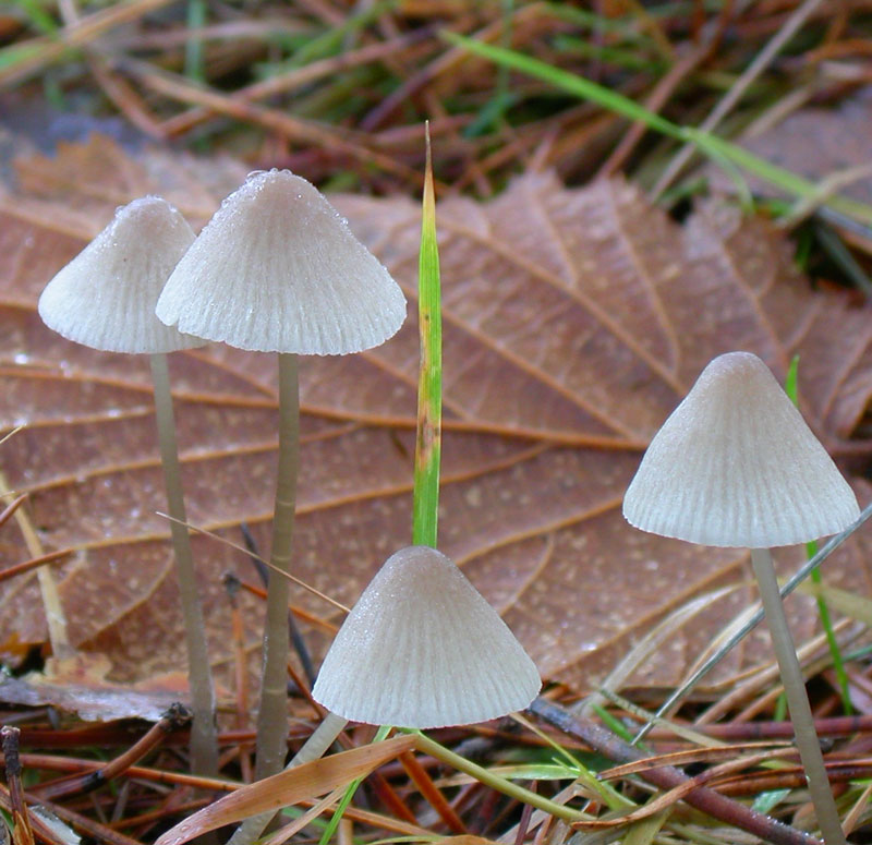
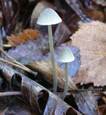
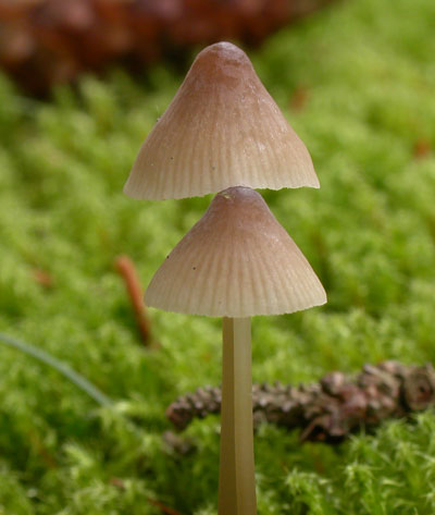
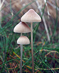
Pileus 5-25
mm across, narrowly to broadly conical or campanulate, sulcate,
translucent-striate, pruinose, glabrescent, fairly dark
brown to pale brown or greyish brown, darker and often with
a pink shade at the centre. Lamellae
13-24 reaching the stipe, ascending, narrowly adnate,
white to pale grey, often turning pale brownish flesh-colour
to dingy pink. Stipe 25-90
x 1-2 mm, hollow, terete, equal, straight to curved, fragile,
pruinose, glabrescent, whitish or grey to grey-brown, usually
darker in lower parts, the base densely covered with long,
white fibrils. Odour of iodine
when drying (best experienced after having been kept in
a box for a while). Taste insignificant.
Basidia
24-35 x 7-10 μm, clavate, 2-spored and 4-spored
Spores
8-12.5(-14.5) x 5-6.3 μm (from 2-spored basidia) or
8.5-11.8 x 4.5-6.5 μm (from 4-spored basidia), Q 1.7-2.5, Qav~2, pip-shaped,
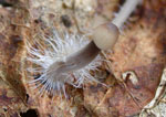
smooth, amyloid. Cheilocystidia
18-76 x 9-27 μm, forming a sterile band, cylindrical,
clavate, obpyriform, obovoid, spheropenduculate, or less
frequently somewhat irregularly shaped, mostly stipitate
(but sometimes sessile), ccovered with mostly evenly
spaced warts or straight to curved or flexuous, cylindrical
excrescences 2-10 x 0.5-1.5 μm. Pleurocystidia similar. Lamellar trama dextrinoid. Hyphae of the pileipellis
2-5.5 μm wide, diverticulate, covered with simple to branched excrescences,
often forming dense, coralloid masses. Hyphae
of the cortical layer of the stipe 2-4.5 μm wide, diverticulate, with excrescences 1-10 x 1-2 μm, terminal cells absent or rare. Clamps present in all tissues, both in 2-spored and 4-spored form.
Microphotos of cheilocystidia
Microphotos of hyphae of the cortical layer of the stipe
When Mycena metata is found in great
numbers under Picea abies, and showing a pink shade
at the centre of the pileus and on the lamellae, identification
seldom causes any trouble. Sometimes the pink shade is quite
distinctive; I have even collected specimens that have been
entirely pink. This species can, however, appear in different
colour varieties, and often there is no trace at all of
a pinkish colour. Then it may be mistaken for Mycena
filopes. This problem was treated
by Maas Geesteranus (1984: 437):
Mycena filopes:
a) Pileus surface giving the impression of being rimose,
with the innate fibrils splitting much in the way of the
superficial fibrils of the pileus of some Inocybe, and b)
imparting a silvery lustre on drying out, c) pileus not
becoming tinged with pink, d) lamellae not or only rarely
turning slightly pinkish, e) sessile cheilocystidia often
more frequent than the stipitate ones, f) stipitate cheilocystidia
up to 30 µm long, g) terminal cells of the stipe cortex
always present, numerous and easy to find.
Mycena metata: a)
Pileus surface either not rimose or without apparent texture,
b) without silvery lustre, c) pileus and / or lamellae usually
becoming tinged with pink, d) stipitate cheilocystidia often
more frequent than the sessile ones, e) stipitate cheilocystidia
often greatly varying in size and the bigger ones usually
more voluminous than their counterparts in M. filopes, reaching
more than 70 µm in length, f) terminal cells of the
stipe cortex absent or, if present, rare and mostly hard
to find.
Because of the great morphological variation, it would be natural to think that more than one species is involved. This should be investigated with molecular methods. So far, preliminary results from the Norwegian Barcoding Project (NorBOL), are not indicating a complex.
Robich (2003) described a number of new species from Italy that would fit in with the M. metata species concept at this web site.
Microscopic illustrations.
Go to sect. Filipedes.
|