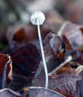
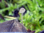
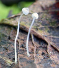
Solitary or in groups on fallen leaves on the ground under Salix spp. (e. g. Salix lapponum), and occasionally on small, partly buried, twigs, in alpine sites. Summer to autumn. Not uncommon in Norway, but probably overlooked because of the tiny size and the unaccessible habitats. Recorded from three counties: Telemark, Buskerud and Hordaland. Reported from Germany (Lehmann & Lüderitz 2018).
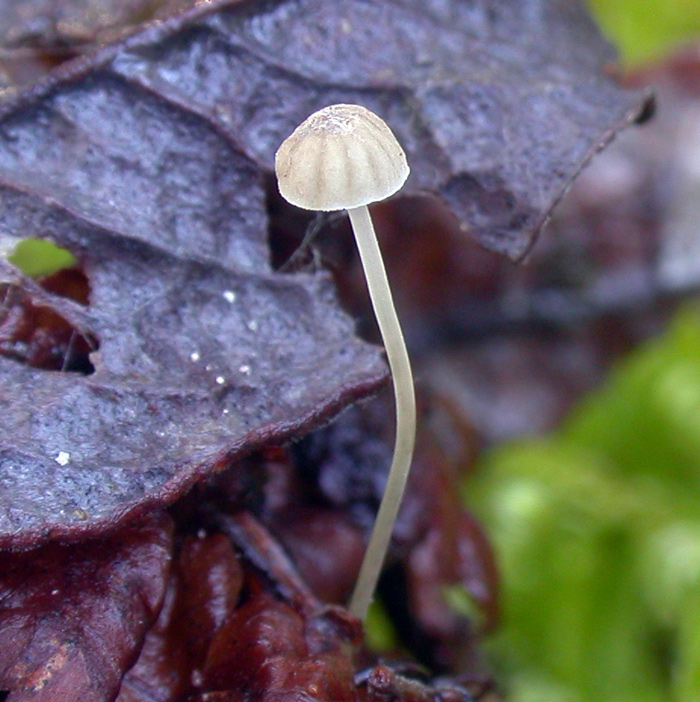
Pileus 1-2 mm across, hemispherical, then conical to parabolical, flattening to convex, then more or less plano-convex with or without a small papilla centrally, occasionally somewhat depressed at the centre, pruinose, glabrescent, shallowly sulcate, hardly translucent-striate, pale brown or pale grey-brown or grey to dark grey, sometimes pale grey, the centre dark brown to dark grey. Lamellae 4-8 reaching the stipe, with or without lamellulae, ascending, usually well developed and fairly broad, but occasionally only showing as faint ridges, the edge concave to convex, narrowly adnate to broadly adnate, sometimes decurrent with a very short tooth, pale brown, pale grey to white, the edge pallid. Stipe 10-30 x 0.1-0.2 mm, very thin, hollow, equal, terete, firm, pruinose, glabrescnt, becoming shiny, curved to flexuous, brownish grey to watery grey becoming more whitish; sometimes dark grey at the apex in younger specimens, often conspicuously insititious but occasionally attached to the substratum by a whorl of radiating, flexuous, white mycelial fibrils. Odour and taste indistinctive.
Basidia 18-33 x 7-11 µm, clavate, (2-) and 4-spored, clamped, with plump sterigmata up to 5 µm long. Spores 7.5-10 x 4.8-6.5 µm, Q 1.3 - 1.9, Qav ~ 1.6, broadly ellipsoid to pip-shaped, smooth, amyloid. Cheilocystidia 21-40 x 6-11 µm, occuring mixed with the basidia, clamped, cylindrical, subclavate, sublageniform, smooth. Pleurocystidia not seen. Lamellar trama dextrinoid, reddish brown in Melzer's reagent. Hyphae of the pileipellis 5-10(-17) µm wide, clamped, densely covered with cylindrical, straight excrescences 1-3.5 x 0.5 µm; terminal cells up to 40 x 15 µm, clavate to subcylindrical, covered with short, straight excrescences. Hyphae of the cortical layer of the stipe 1-7 µm wide, clamped, densely covered with cylindrical, sometimes more thorn-like, straight excrescences 1-6 x 0.5-1 µm; terminal cells not typically differentiated but a few observed at the base of the stipe being clavate, 26-35 x 5-10 µm, densely covered with cylindrical excrescences 1-4 x 0.5-1 µm.
Microphoto of the cheilocystidia 1
Microphotos of the cheilocystidia 2
Microphotos of the cheilocystidia 3
Microphotos of cheilocystidia, pileipellis and stipitipellis
Microphotos of basidia, spores and hyphae of the pileipellis
Microphotos of spores
M. guldeniana is a member of sect. Polyadelphia, where it shares some essential features with Mycena terena. Both species are associated with Salix, and both species have smooth cheilocystidia, an uncommon feature in the section. Aronsen & Perry (2012) could, based on molecular analyses, differentiate the two species. Recent sequences obtained by the Norwegian Barcoding Project (NorBOL), however, show no difference between the two species. Until more collections have been studied, I prefer to regard them as two separate species.
There are several morphologic features that can be used to distinguish the two taxa.
The differences are tabulated below:
| |
Mycena guldeniana |
Mycena terena |
| Pileus |
pale brown / grey-brown |
pale beige or very pale grey, soon fading to white |
| Spores |
Qav ~ 1.6 |
Qav ~ 1.8 |
| Hyphae of the pileipellis |
with prominent terminal cells |
without prominent terminal cells |
| Habitat |
on fallen leaves and twigs under Salix spp. in alpine area |
on fallen leaves of Salix caprea in coastal lowland |
Gulden & Jenssen (1982) reported on an unknown species of Mycena, collected on a dead leaf at Nordnut, Finse, Hordaland 11 Aug. 1979. Their material was "too scanty for a formal description of a new species". The description, however, indicates that their unknown species was identical with the present taxon. |