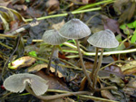
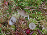
Scattered on debris of various deciduous trees as well as on fallen needles of coniferous trees (e. g. Juniperus and Picea), also on turfs and among Sphagnum. Summer to early winter. Very common and widely distributed all over the area, also present in alpine Salix shrubs. In Norway one of the most common species and can be found all over the coutry.
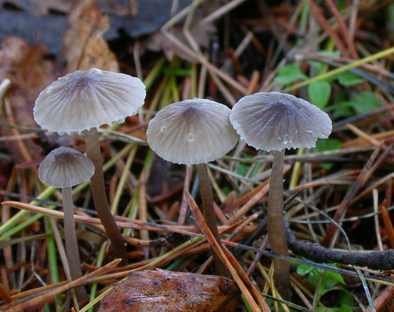
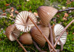
Pileus 10-25 mm across, conical to campanulate, more or less umbonate, somewhat flattening with age, the margin sometimes flaring and upturned with age, sulcate, translucent-striate, pruinose, glabrescent, somewhat lubricous, grey, grey-brown to fairly dark sepia brown, darker at the centre, the margin paler. Lamellae 13-18(-23) reaching the stipe, ascending, narrowly to somewhat broader adnate, sometimes decurrent with a short tooth, smooth to veined, becoming dorsally intervenose with age, at first whitish, then brownish white to pale grey-brown, edge whitish. Stipe 50-85 x 1-3 mm, hollow, somewhat elastic, equal or somewhat broadening downward, terete, straight to curved, somewhat pruinose all over, glabrescent for the greater part, exuding a milk white fluid when incised or broken, grey-brown, paler above, darker below, the base densely covered with long, coarse, white fibrils. Odour indistinctive, of earth or raphanoid. Taste mild.
Basidia 25-36(-49) x 7-9 µm, clavate, 4-spored, with sterigmata 6-8(-12) µm long. Spores 10-14 x 5-6 µm, Q 1.9-2.6, Qav~2.1, elongated pip-shaped to almost cylindrical, smooth, amyloid. Cheilocystidia 39-95 x 8-18 µm, occuring mixed with basidia or locally forming a sterile band, generally fusiform but also clavate to obovoid, simple to furcated, more rarely with coarse, lateral or apical excrescences. Pleurocystidia fusiform. Lamellar trama dextrinoid. Hyphae of the pileipellis 1-3.5 µm wide, branched, sparsely to densely covered with excrescences 2-4.5 x 1-2 µm, tending to become somewhat gelatinized. Hyphae of the cortical layer of the stipe 1.5-4.5 µm wide, covered with widely spaced to more crowded, simple to furcated excrescences 1.5-10 x 1-2 µm, the terminal cells smooth to diverticulate, up to 4.5 µm wide. Clamp connections present in all tissues.
Microphotos of cheilocystidia
Microphotos of caulocystidia
Mycena galopus is easily identified on account of the milk white fluid in the stipe. Sometimes, in dry specimens the fluid may not be visible and then it is not easy in the field to separate from other grey-brown species. In these cases it can be identified by microscopic features as the large, narrow spores and large, fusiform cheilo- and pleurocystidia.
Three varieties of this species, all with white fluid in the stipe: galopus, candida and leucogala, were recognized by Pearson (1955). The last variety was subsequently classified as a separate species by Dennis, Orton & Hora (1960). Maas Geesteranus (1988 d) treated M. leucogala as a species in its own right and provided some arguments that seem to be rather weak. Forms intermediate between the typical black fruitbodies of M. leucogala on burnt ground and those of the grey-brown or pure white varieties of M. galopus are common, and mycelia of the three taxa appear to be serologically identical (Chard et al. 1983), but interbreeding has not been confirmed (Frankland 1984).
|