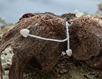
Scattered or gregarious on nuts and husks (seldom on leaves) of Corylus avellana from May to the end of October. Known only from type locality, in the Netherlands.
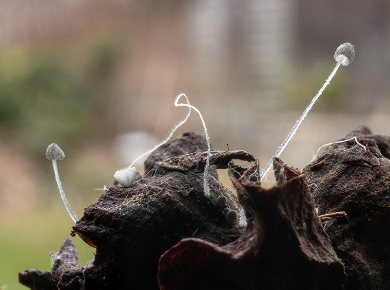
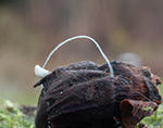
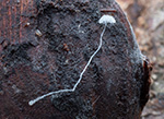
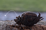
Pileus up to 2(-2,3) mm diam., initially paraboloid, expanding to obtusely conic or broadly convex in age; young covered all over with groups of grey to dark grey (sometimes nearly blackish grey) ‘sugar-like’ granules on a pale grey surface, mature white, still bearing small amounts of granules especially in the centre, translucent-striate, margin crenulate, old very thin-fleshed, translucent. Lamellae subventricose to ventricose, adnexed to narrowly adnate, with lamellulae of variable length, (6-)8-10 reaching the stipe, white. Stipe 8–20 x 0.08-0.2 mm, central, filiform, hirsute, watery grey to watery white, at the base slightly inflated, with a small basal disc, not always easily seen and sometimes apparently absent; disc margin granulose. Primordia hemisphaerical to paraboloid, covered with dark grey granules, initially forming a closed layer, soon breaking up to a non-contiguous layer upon a light grey surface, more whitish at the margin; dried dark grey. Smell and taste insignificant.
Basidia 9–20 x 7–10 µm, short clavate to obpyriform, 4-spored, rarely 2-spored, sterigmata 2–4 µm long. Spores [100/5] 7.3-8.7-10.2 x 3.9-4.7-5.5 µm, Q = 1.54–1.87–2.19, Qav = 1.81-1.91, ellipsoid, smooth, hyaline, thin-walled, amyloid. Cheilocystidia very sparse or absent, 8–24 x 5.5–13 µm, clavate to broadly clavate, sparsely spinulose in the upper part but more densely spinulose near the pileus margin; spinulae 0.5–1 x 0.5 µm, cylindrical to subconical. Pleurocystidia absent. Pileipellis a cutis with acanthocysts and cherocytes; hyphae 1.5–13 µm diam., spinulose or smooth, dextrinoid. Central acanthocysts globose, subglobose, thin-walled, 8–22 x 9–19.5 μm, densely spinulose, grey brown, grey or hyaline, inamyloid, sometimes seen originating from septate, thin-walled, smooth or spinulose hyphae of about 3 µm diam.; spinulae 0.5–1 x 0.5–1.5 μm, cylindric or subconical. Marginal acanthocysts clavate, broadly clavate, ovoid or subglobose, thin-walled, 11–28 x 8–13 μm, densely spinulose, grey or hyaline, inamyloid, spinulae 0.5–1 x 0.5–1.5 μm, cylindric or subconical. Cherocytes roundish to obtuse angular, with 2–6(7) obtuse or obtusely conical lobes and with dark grey brown vacuolar contents, covered with spinulae and warts; 9–28 x 9–25 µm (disregarding lobes), thick-walled, walls grey or hyaline, up to 5 µm, lobes extending up to 11 µm, warts hemisphaerical, 0.5–2.5 x 0.5–3 µm, often seen originating from a septate, slightly thick-walled, smooth or spinulose hyphae of about 3 µm diam., content slightly dextrinoid. Hypodermium composed of inflated hyphae up to 28 μm diam, dextrinoid. Lamellar trama composed of inflated hyphae up to 18 µm, dextrinoid. Cortical and medullary hyphae of the stipe 3-19 μm diam., parallel, smooth, dextrinoid. Basal disc cystidia globose to subglobose, spinulose, connected to the stipe base by a dense layer of short smooth or spinulose cells from about 3-5 x 6 µm, partly loosening with age; 9–23 x 6–20 µm diam., grey or hyaline, inamyloid; spinulae 0.5–1 x 0.5 µm, cylindrical, conical or subconical. Caulocystidia abundant, 9–142 x 4.5–16 μm, hyaline, thin-walled, inamyloid, ranging from short and broadly clavate or subcylindric to long and cylindric, apex even or somewhat wider, obtuse, densely and evenly spinulose overall, spinulae 0.5–1(2) x 0.5–1 μm, cylindric or subconical. Clamps present but very rare, only seen at the base of a few basidia.
Microphotos of cherocytes, acanthocysts and cheilocystidia
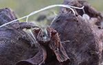
The description has been copied from the type description (Jagers et al. 2023). I received a number of hazel nuts from the Netherlands and put them in a small plastic box with moist conditions. After a few weeks the first fruitbodies appeared, and the fungus continued producing fruitbodies for about two months. This made it possible to study the species in all stages of age and to study the microscopical details too. Many thanks to Marian Jagers.
Mycena amoena belongs to Mycena sect. Amparoina T. Bau & Q. Na. (See Jagers et al. 2023 for a further discussion.) This section contains species primarely distributed in tropic habitats and was until recently not recorded in Europe. The phylogenetically closest related species to M. amoena are M. melanovelis Traba, Couceiro & M. Villarreal nom. prov. (Traba-Velay et al. 2021) and M. lasiopus BAP635 (and BAP603, Cooper et al., 2018).
M. melanovelis nom. prov., also has cherocytes with coloured vacuolar contents, but contrary to M. amoena, its cherocytes have small spines instead of lobes. It also differs from M. amoena by lacking a basal disc, having bigger basidiomata, smaller spores, and cheilocystidia forming a sterile band. M. lasiopus differs from M. amoena in having a somewhat larger, rather dark pileus, a radially lamellate basal disc, clamps in all tissues, broader spores, numerous cheilocystidia, and its caulocystidia are almost smooth at the apex.
|