Growing singly or gregarious on small twigs and other terrestrial plant debris, often apparently growing on the ground, under Salix lapponum, and probably under other Salix species, and in Betula forests in alpine areas, but in Finland also in “alpine” pine-spruce heath forest. Autumn. Rare, in Europe only recorded from Norway, Sweden and Finland.
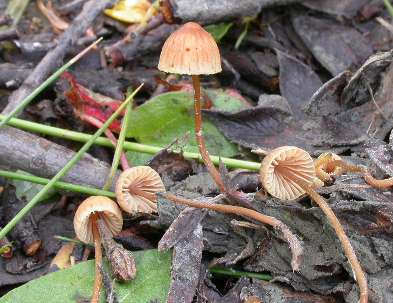
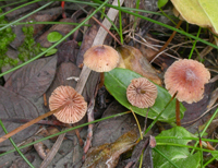
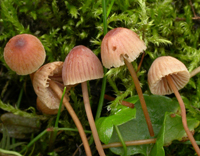
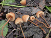
Pileus 4-13 mm across, acutely
conical in young stages, becoming broadly conical with age,
sometimes with a small umbo or papilla, sulcate, translucent-striate
or not striate, pruinose, glabrescent, fulvous, usually
with a dark yellow-brown centre, somewhat paler at the margin,
drying to yellowish brown; sometimes with a pink hue. Odour
of iodoform when drying. Lamellae
13-16 reaching the stipe, ascending, narrowly adnate,
not decurrent with a tooth, yellowish to pale fulvous, sometimes
with a pink tinge. Stipe 25-65 x 0.5-1 mm, somewhat firm, hollow, terete, equal, straight
to somewhat flexuous, pruinose, glabrescent, fulvous to
brownish, often fairly dark, the base densely covered with
long, coarse, flexuous, whitish fibrils.
Basidia
22-30 x 5.5-7.5 µm, slenderly clavate, 4-spored,
with sterigmata up to 5 µm long. Spores
7.5-12 x 4.3-5.5 µm, pip-shaped to broadly pip-shaped, Q 1.6-2.5, Qav ~ 2.0, smooth,
amyloid. Cheilocystidia
14-32 x 6-14 µm, forming a sterile band, clavate, covered with more or less evenly spaced warts and
cylindrical, simple to branched excrescences 1-12 x 0.5-1 µm. Pleurocystidia
absent. Lamellar trama dextrinoid.
Hyphae of the pileipellis
1-5 µm wide, covered with warts and simple
to branched, straight to curved cylindrical excrescences,
tending to become somewhat gelatinized, and eventually forming
dense masses. Hyphae
of the cortical layer of the stipe 1.5-4
µm wide, not gelatinized, covered with warts
or straight to somewhat curved cylindrical to conical excrescences
2-6 x 1 µm, terminal
cells cylindrical to clavate, diverticulate. Clamp connections present in all tissues.
Microphotos of cheilocystidia, hyphae of the pileipellis and hyphae of the cortical layer of the stipe.
Microphotos of cheilocystidia and hyphae of the pileipellis.
The fulvous or tawny to ochraceous colours, usually present in all parts of the fruitbodies are very peculiar for a Mycena. This species is strikingly similar to a Galerina, and the rather few records may be due to the possibility that it has been overlooked because of its occurrence in habitats with many Galerina species. In the field it can be distinguished from a Galerina on account of the paler gills.
In sect. Filipedes, M. alexandri belongs to a group of species with a smell of iodoform when drying out, more easily recognized after it has been kept in a box. It differs, however, from all other species in the section by the colours.
Aronsen & Gulden (2007) proposed M. aphanes to accommodate European material and claimed it to be different from the North American M. alexandri. DNA sequences performed recently, however, indicate that the two taxa are conspecific (Aronsen & Larsson 2016).
Go to sect. Filipedes
|