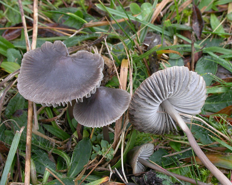
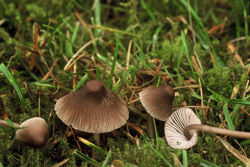
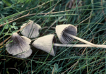
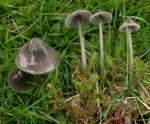
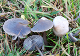
On lawns, meadows, and grassy areas. Autumn. Common. Widely
distributed in Norway, but not many records.
Pileus 10-35
mm across, conical with acutely pointed centre, campanulate
with or without an umbo, convex to more flattened with a prominent,
broad umbo, pruinose, glabrescent, translucent-striate when
moist, sulcate, somewhat lubricous when moist, hygrophanous,
black to dark brown in
the centre, grey-brown to grey towards the grey to whitish margin, fading to dark grey to grey. Lamellae
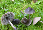
17-25 reaching the stipe, ascending, narrowly adnate,
sometimes with a short tooth, dorsally intervenose with
age, dark to pale grey with the edge paler. Stipe
25-70 x 1.5-5 mm, hollow, straight or somewhat curved, fragile, equal or somewhat thicker towards the base, terete, with age sometimes somewhat depressed and fissured,
pruinose at the apex, glabrous below, pale brown, grey-brown or greyish,
darker below, the lower parts sometimes blackish brown, the base
covered with long, coarse, flexuous white fibrils. Odour
indistinctive, acidulous or somewhat raphanoid when cut, not nitrous.
Basidia 27-36 x
7-10 µm, clavate, 4-spored. Spores 7-11 x 5–6.5 µm, Q 1.5-2.1, qav ~ 1.7, pip-shaped, smooth, amyloid. Cheilocystidia
25-75 x 6.5-22.5 µm, fusiform, lageniform,
subcylindrical or somewhat irregularly shaped, apically
with a simple or furcate neck, sometimes also with few,
very coarse excrescences. Pleurocystidia
scarse, fusiform. Lamellar trama dextrinoid. Hyphae of the pileipellis
2-4.5 µm wide, covered with simple to very much branched, cylindrical
excrescences, which tend to form dense masses and become
gelatinized. Hyphae of the cortical layer of
the stipe 1-3.5 µm wide, diverticulate, with terminal cells often hard to find, variously diverticulate. Clamp connections present at all tissues.
Mycena aetites
is a member of sect. Fragilipedes,
and is not always easy to identify. It can be
found together withseveral other members of the section. M.
leptocephala has a nitrous smell, smooth hyphae of the stipe cortex and typical,
inflated terminal cells. M.
parca also has smooth stipitipellis hyphae, and the cheilocystidia are more
homogeneously lageniform. M. austera has a nitrous smell, smooth to sparsely diverticulate stipitipelliis, and large, conspicuous terminal cells in the pileipellis. M.
aronsenii can be distinguished on account of the smaller spores, the hyphae of
the cortical layer of the stipe which are covered with
more or less curved to coiled excrescences and the coarsely
diverticulate terminal cells. Mycena ustalis has smooth hyphae of the pileipellis, and M. cretata has long, hair-like caulocystidia. They both have a nitrous smell. |
|
Another species looking
quite similar to M. aetites, is M.
abramsii. It usually
grows on wood, and the cheilocystidia tend to have a conspicuously
acute neck.
Maas geesteranus (1988) gave a description of M. murina (Murrill) Murrill, a species known from both Europe and USA. He referred to a Swedish collection (as M. stannea (Fr.) Quél.in Lundell & Nannfeldt 1935). I do not know this species. The cap was aid to be grey with a faint bluish tint. The microscopic features seem to match M. aetites. Ludwig (2012) suggested that M. tristis is conspecific with M. aetites. They, admittedly, look rather similar but are treated as two separate species here.
|